
Gel electrophoresis is a commonly used technique in biology for separating nucleic acids based on their charge and size. This technique is essential for many biological experiments.
Scientists often work with DNA, extracting it from various samples, manipulating it, joining it, cutting it into smaller fragments, and using enzymes, among other things. However, before they can perform these techniques, they need to be able to visualize the DNA accurately.
Have you ever wondered how scientists can see the tiny DNA molecule? Well, the answer is “electrophoresis”.
Electrophoresis is a method used to separate charged biomolecules, such as DNA, RNA, and proteins, using electricity. Arne Tiselius first used electrophoresis to separate biological molecules in 1931. Even after almost a century, electrophoresis remains an important technique and the only way to separate complex biomolecules.
What Is The Principle Behind This Technique?
The main principle behind electrophoresis is the use of electricity to separate biomolecules based on their charge and size. The separation is based on the fact that “like charges repel each other and opposite charges attract each other”.
Most biomolecules carry a charge, either positive or negative. DNA and RNA have a negative charge due to the phosphate groups in their structure, while proteins can be either positive or negative, depending on the amino acids they are composed of.
When this separation is performed on a jelly-like medium, typically made of agarose (a carbohydrate polymer), it is called agarose gel electrophoresis.
What Is The Gel In Gel Electrophoresis?
The gel in gel electrophoresis refers to the matrix on which the biomolecules are separated. It is a clear and jelly-like substance, similar to the gelatin we consume as food.
The most commonly used matrix for electrophoresis is agarose, a polysaccharide. Agarose is chosen for two reasons. First, it can form a mesh or crosslink, which acts as a sieve through which biomolecules can travel based on their size. Second, agarose is neutral, meaning it does not react with the biomolecules being separated, reducing the chances of obtaining false results.
Let’s examine the setup of gel electrophoresis to understand how the separation occurs.
 The diagram illustrates an electrophoresis system with a positively charged anode and a negatively charged cathode immersed in an electrolyte (buffer) solution. The sample wells are used to load the biological samples to be separated. (Photo Credit: M. PATTHAWEE/Shutterstock)
The system consists of a small tank with two electrodes—an anode (positively charged) and a cathode (negatively charged)—submerged in a buffer solution. This buffer solution is often an electrolyte, a solution capable of conducting electricity.
The gel is placed in the buffer solution and contains small wells where the samples can be loaded. The sample wells are positioned close to the negative electrode, causing the negatively charged samples to move towards the positive electrode when the power supply is turned on.
The repeating unit of agarose consists of D-galactose and 3,6-anhydro L-galactose. The movement of biomolecules in the gel depends on their charge and size. Biomolecules with a negative charge move towards the positive terminal and vice versa. Smaller fragments, which are lighter and have low molecular weight, can travel quickly through the gel and cover a larger distance. On the other hand, larger fragments with higher molecular weight move slowly due to the pore size of agarose. The rate of movement also depends on the percentage of agarose used in the gel. Low-percentage agarose gels have larger pore sizes to allow faster movement of larger molecules, while higher percentage gels with smaller pore sizes are better for separating smaller DNA molecules. A diagram illustrates how molecules of different sizes move through the agarose gel, with smaller molecules traveling a greater distance than larger ones.
Methods of Visualizing DNA
When examining the electrophoresis buffer, agarose gel, or DNA samples, they are all transparent. So how can we visualize the DNA? We utilize a colored dye that attaches itself to the DNA or biomolecules. The most commonly used dye for binding to DNA is ethidium bromide (EtBr).
The dye can be added to either the agarose gel or the buffer that fills the apparatus. In either case, it needs to come into contact with the DNA and bind to it.
Once the dye has been added, the gel is exposed to ultraviolet (UV) light. Under UV light, ethidium bromide emits a bright light. Since ethidium is already bound to the DNA, the areas on the gel that appear bright indicate the presence of DNA.
When working with EtBr, it is important to exercise caution as it is known to cause cancer (carcinogenic). Proper handling and disposal are necessary.
The DNA will appear as bright, vertical lines known as bands against a dark background. Each band consists of DNA molecules of the same size. Intact bands indicate high-quality DNA. If the DNA is of low quality or contains contaminants, the DNA band will not be intact and will spread out, resulting in a smear.
If we wish to determine the size or molecular weight of the DNA, we can use DNA ladders. These are commercially available DNA samples with known sizes. By loading the DNA ladder alongside the sample DNA (whose molecular weight is unknown), we can compare the distance migrated by both DNA samples to determine the molecular weight of the sample DNA.
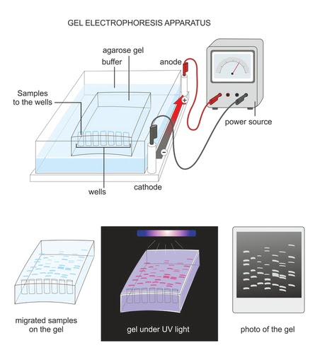 The image illustrates the complete process of agarose gel electrophoresis. The samples are placed in the wells of an agarose gel immersed in buffer and connected to a power supply. After the sample has migrated through the gel, it is visualized under UV light to assess the quality and quantity of DNA. (Photo Credit: Soleil Nordic/Shutterstock)
What Can Affect the Movement of DNA in Gel?
There are several factors that can influence the way DNA migrates in a gel. The concentration of agarose used has an inverse relationship with the size of the DNA molecule; larger DNA requires lower concentrations of agarose and vice versa.
The voltage applied also plays a role. Higher voltage leads to faster movement of the samples, but the DNA bands may not remain intact. Lower voltage results in slower DNA migration, but the bands will be distinct. Therefore, a compromise may be necessary between time and voltage for proper DNA separation.
The presence of ethidium bromide (EtBr), the quality of agarose, and the pH and composition of the buffer used also affect DNA mobility.
A Final Thought
Agarose gel electrophoresis is the primary and perhaps the only technique for determining the quality and size of nucleic acids in various areas of biological research. The past two decades have witnessed significant advancements and expansion in the application of this technique.